
The Lundberg Institute
  • Our Mission
  • Dedication
  • Our Platform
  • Who We Are
  • Contact
  • Resources
  • Press
  • Curious Dr. George Blog
  • Search
  • Menu Menu
Curious Dr. George | Plumbing the Core and Nibbling at the Margins of Cancer

Capturing Patients’ Real-World Experiences to Improve Cancer Research and Care

February 18, 2019/in CDG at TLI

A Q&A with Grace Castillo-Soyao, founder and CEO of Self Care Catalysts; grace@selfcarecatalysts.com
Q: You are well known as a visionary in the field of Real World Experience-Evidence (RWEE). As the founder and CEO of Self Care Catalysts, headquartered in Toronto, how do you see RWEE evolving to favorably impact the field of oncology?
A: I started Self Care Catalysts with some very basic questions. Why is the patient at the farthest end of the care line, treated as simply the recipient of care? It’s a very industrial mindset, a bit like an assembly line; the patient as something to be acted upon. But patients are often experts at their own conditions, including the many kinds of cancer. Why are they not invited to become participants in their own care, in contributing their own experiences? Why are patient experiences not considered to be scientifically valid?
As noted physician Sir William Osler famously said, “It is much more important to know what sort of a patient has a disease than what sort of a disease a patient has.”
About 90% of disease management happens at home, but very little information has been gathered about what happens in between clinical visits, and how self care might impact outcomes. This knowledge gap between caring for and living with cancer is wide, yet patients’ experiences—such as the burden of their cancer, their treatments, side effects, and the impact on their sleep, diet, relationships, and work—are barely acknowledged, documented, or discussed with their clinical care team. It’s not because nobody cares; it’s because of a time barrier and an economic barrier. Physicians traditionally get paid for services that happen inside the clinic, not beyond it.
Oncology is one of the disease states for which treatment technologies have undergone significant growth and evolution. Recently, the U.S. Food and Drug Administration has been employing new approaches to expedite the drug development and approval process. These include an emphasis on understanding the “real world,” or what happens with patients outside of clinics and clinical trials.
Because of these changes, and the complexity of cancer management by patients themselves, clinical trials will increasingly need to employ new measures that account for patients’ day-to-day experiences with their disease and treatments. And, as more promising cancer drugs enter the oncology market, the need to collect real world patient experience evidence (RWEE) will become a cornerstone in patient care and treatment decisions.
This shift in oncology will be enabled by the use of digital tools that collect data in a more rigorous way. Such tools can now address long-standing doubts and concerns regarding the validity and subjectivity of data reported by patients. The same tools can also be applied to disease management, which we at Self Care Catalysts believe will dramatically improve clinical outcomes. Indeed, Ethan Basch, MD, MSc, an oncologist at the University of North Carolina and one of our advisors, recently demonstrated that enabling patients to report their symptoms in clinical cancer care improved outcomes dramatically.
For the medical and scientific community, I’m sure it’s comforting to see that collecting patient-reported outcomes in cancer actually provides a dramatic improvement to care! We expect to see that digital engagement has a dramatic effect in how well a patient’s disease is managed and the quality of outcomes that can be gained, and we’ll see it in ever more rigorous studies.
At Self Care Catalysts, we design and build tools to capture the true patient experience. Patient experience data from approximately 3,500 cancer patients who use our Health Storylines Real World Experience Evidence platform reveal that their most common concerns are related to psychosocial, mood, quality-of-life, disease, and treatment experiences. These factors are hardly captured in clinical, lab, and medical reports at all, and not accurately represented in validated quality-of-life surveys.
RWEE combined with traditional clinical, medical, and even genomics data will bring quality of life realities to oncology, and it will advance many aspects of research and care, including drug development, clinical trials, and commercialization. Most importantly, RWEE will improve how cancer patients are involved in their own care, leading to big improvements in outcomes.
***
Copyright: This is an open-access article distributed under the terms of the Creative Commons Attribution License, which permits unrestricted use, distribution, and reproduction in any medium, provided the original author and source are credited.
https://lundberginstitute.org/wp-content/uploads/2018/01/cdg-speech-bubble-trimmed-300-1.png 300 300 melindaroberts https://lundberginstitute.org/wp-content/uploads/2020/08/Artboard-1.png melindaroberts2019-02-18 13:19:102019-08-14 17:10:36Capturing Patients’ Real-World Experiences to Improve Cancer Research and Care
Curious Dr. George | Plumbing the Core and Nibbling at the Margins of Cancer

The Crucial 90% Missed by Doctors on Computers

February 11, 2019/in CDG at TLI
A Q&A with Kevin B. Knopf MD, MPH, chairman of hematology and oncology at Highland Hospital in Oakland, California; kevinbknopf@gmail.com
Q: A successful patient-physician relationship depends upon effective bidirectional attention and mutual understanding. Many patients and physicians believe that common current versions of mandated electronic health records (EHRs) severely impede that interaction, especially eye contact. How can a competent and caring clinical oncologist overcome that problem?
A: For all my faults as a doctor, and I’m sure there are many, there is one thing I think I do correctly, and that is I am never on a computer in front of a patient.
I hear from many colleagues that they can be efficient and personable while going back and forth from the patient to their electronic health record (EHR)—and it is true there are various levels of skill here. However, none do as well, in my opinion, as a computer-free patient environment. I say this from my side as a patient having seen dozens of doctors myself—nothing compares to a doctor who spends all of their time looking you in the eye and interacting face to face. This human contact costs nothing, and yet is so vital.
I’m hardly a Luddite; from my side as a doctor the math is infallible here: approximately 90% of all human communication is nonverbal, so if I am typing on a computer I’m only processing 10% of what is going on and my clinical efficiency drops accordingly. I’m convinced that this 90% is important not just for healing the patient; the way questions are answered and interpreted subtly can allow me to order fewer unnecessary tests and be a more cost-effective doctor.
In oncology many of the discussions veer into existential and religious domains, and this can’t be done except on a very intimate level. My greatest enjoyment in clinic are my patient interactions; for my own wellbeing, I hate to poison these with the EHR.
Some days it seems that the patient note in the EHR deteriorates to a “set of lies agreed upon,” particularly the review of symptoms and the physical exam, which is often templated and describes some Faulknerian version of the truth of what is actually happening.
In today’s world of “high throughput healthcare” designed to maximize relative value units (RVUs) and document in the EHR to upcode, it is the patient who suffers. And yet, it is the patient for whom we all go into medicine. I have done my own time and motion studies on this topic, and my current habit of reviewing the EHR in a separate room before I see the patient and then returning to that room to write the encounter after is more efficient than trying to go back and forth between the patient and the computer.
As a third-year medical student, now 30 years ago, the attending asked me to perform a history and physical on an inpatient in front of the entire team (gulp). I’ll never forget that afterwards he said, “you did a good job, but I want you to pull up a chair next time and sit down when you are talking to the patient.” This echoes the saying of the great oncologist Dr. Jamie Von Roenn: “Don’t just do something, sit there.” Indeed, there is a pleasure in the slowness of the patient encounter that most of us went into medicine to experience—and the EHR continually interferes with that process.
I recently spent about 10 days as a patient in the hospital—in three different hospitals—and different health care providers spent more or less time on a computer in front of me. The best came in my room and were computer free. In the end only one pulled up a chair and sat down to ask how I was doing—my friend the breast surgeon, who is a generation younger than me. I notice in her clinic she is an “EHR-free doctor” as well. Consider this a start.
***
Copyright: This is an open-access article distributed under the terms of the Creative Commons Attribution License, which permits unrestricted use, distribution, and reproduction in any medium, provided the original author and source are credited.
https://lundberginstitute.org/wp-content/uploads/2018/01/cdg-speech-bubble-trimmed-300-1.png 300 300 melindaroberts https://lundberginstitute.org/wp-content/uploads/2020/08/Artboard-1.png melindaroberts2019-02-11 13:15:572019-08-14 17:13:23The Crucial 90% Missed by Doctors on Computers
Curious Dr. George | Plumbing the Core and Nibbling at the Margins of Cancer

The Promise of Plerixafor in Glioblastoma Treatment

November 13, 2018/in CDG at TLI
A Q&A with Adan Rios, MD; Professor in the Division of Oncology-Department of Internal Medicine of The University of Texas McGovern Medical School at Houston, Texas Medical Center; adan.rios@uth.tmc.edu
Q: Glioblastoma multiforme (GBM) remains a scourge with a typically rapid fatal course resistant to most therapy. All solid tumors must receive sufficient blood supply to grow. Plerixafor is an FDA-approved drug that may inhibit tumor angiogenesis. How might plerixafor be sensibly used off-label as an adjunctive therapy for GBM?
A: Glioblastoma multiforme (GBM) is a CNS (central nervous system) tumor with post-therapy median time to progression of 7 months and median overall survival of 15 months. I decided to use plerixafor for the prevention of recurrence of GBM in one patient treated with standard chemo-radiotherapy five years ago and since then have studied this patient and this subject in depth.
The decision to recommend plerixafor to this patient was borne out from the availability of a human xenograft mice model of GBM developed at Stanford University and in which a variety of experimental conditions clearly demonstrated clinical impact on survival by using plerixafor as an adjuvant to treatment of the experimental GBM with radiotherapy and temodar. An interesting aspect of this model was that plerixafor, on its own, did not have direct anti-tumor effect. This lack of plerixafor anti-tumor effect in the absence of effective established therapy may explain the failure of clinical Phase I-II trials of plerixafor alone in patients with relapsed GBM. In these trials plerixafor was administered intravenously in escalating doses and for periods of up to one month, undoubtedly reaching levels that would have demonstrated direct anti-tumor activity against GBM, should plerixafor have such activity.
It must also be pointed out that the mechanism invoked for the activity against GBM in the Stanford experimental model has less to do with the anti-angiogenic characteristics of plerixafor and expression of CXCR4 receptors by the tumor and endothelial cells and more to do with the active blocking of bone marrow-derived myeloid precursors (BMDCs, MMP-9 expressing CD11b+ myelomonocytes). In the presence of active chemo-radiotherapy against GBM, this active blocking rescues the tumor from the effects of treatment by a process known as vasculogenesis, in opposition to primary angiogenesis. Chemo-radiotherapy eliminates the endothelial cells of the tumor (responsible for primary angiogenesis) resulting in an increased production of hypoxia-dependent HIF-1 and an increased secretion of SDF-1 (CXCL12), the cognate ligand of the CXCR4 receptor, by the tumor and stromal tumor-bed cells. This causes a specific increase mobilization and retention in the tumor bed of BMDCs initiating the tumor recurrence through vasculogenesis. This SDF-1 effect (hypoxia-dependent) can be blocked by plerixafor, an inhibitor of SDF-1-CXCR4 interactions.
The first patient I treated with plerixafor had a relapse of GBM after being free of disease for five years and one month. The small area of relapse was treated with gamma knife and initiation of avastin combined with plerixafor, with no change in his performance status or quality of life. Three other patients of mine are on adjuvant plerixafor off label. One had an early relapse resected and is now back on temodar, an “electric cap,” and continuation of plerixafor. The other two are about to complete one year on adjuvant plerixafor, and while is too early to comment further, they have experienced no unexpected events.
Stanford University’s clinical trials with plerixafor as an adjuvant to chemo-radiotherapy have had ambiguous results that were not as dramatic as expected from the experimental model. I have attributed this to the plerixafor schedule used at Stanford; they administered plerixafor in high doses, but only for the duration of the initial chemo-radiotherapy, and then stopped it. In contrast, the approach I have used is to administer plerixafor on weekly doses until the patient overcomes two critical thresholds: the 7 months of median for progression and the 15 months of overall survival. Plerixafor is long-acting and thus intense daily administration for a relatively short period of time may not be the best strategy of administration for this purpose.
Finally, new data indicates that neural stem cells (NSCs) seem to originate from a very specific zone in the brain, anatomically identified as the sub-ventricular zone (SVZ), from where these cells migrate to different areas of the brain and become transformed into GBM. This raises the fascinating possibility that, regardless of the location of the diagnosed GBM, the prospective irradiation of this specific zone may be of great relevance in the treatment of GBM. This is important because most treatments of GBM with radiotherapy are circumscribed to only the tumor-involved fields.
In conclusion, I believe that there is sufficient experimental evidence to suggest that plerixafor can play a critical role in enhancing the outcomes of conventional therapy of GBM with chemo-radiotherapy. It must be used for a long-enough time to allow the chemo-radio-therapy to eliminate the tumor by a hypoxia-mediated mechanism (elimination of primary angiogenesis) and the blocking of BMDCs, CD11b+ mylomonocytes responsible for vasculogenesis. The outsourcing of GBM stem cells from a specific zone of the brain (the SVZ) is a new and critical observation, the study of which could play a significant role in further understanding the pathogenesis of this tumor and of important changes in our approaches to its therapy.
Copyright: This is an open-access article distributed under the terms of the Creative Commons Attribution License, which permits unrestricted use, distribution, and reproduction in any medium, provided the original author and source are credited.
Tags: BGM, brain cancer, brain tumor, glioblastoma, neural stem cells, Optune, plerixafor, Temodar
https://lundberginstitute.org/wp-content/uploads/2018/01/cdg-speech-bubble-trimmed-300-1.png 300 300 melindaroberts https://lundberginstitute.org/wp-content/uploads/2020/08/Artboard-1.png melindaroberts2018-11-13 17:19:452019-08-14 17:11:47The Promise of Plerixafor in Glioblastoma Treatment
Curious Dr. George | Plumbing the Core and Nibbling at the Margins of Cancer

Might Cancer Be a Metabolic Disease?

October 31, 2018/in CDG at TLI

Thomas SeyfriedBiology Departmentscience

A Q&A with Thomas N. Seyfried, PhD, Professor of Biology, Boston College
Q: As a geneticist, you know that the genomic makeup of cancers recently has captivated much of the scientific community with new knowledge and new treatments. And yet, cancer outcomes remain dismal for many patients. You have written about cancer from a very different perspective. Why do you consider cancer to be a metabolic disease, and how might we look at different therapeutic options under that rubric?
A: Over 1,600 people die each day from cancer in the U.S., according to recent data from the American Cancer Society (Siegel et al., 2018). The failure to manage cancer has been due in large part to the dogmatic belief that cancer is a constellation of genetic diseases. Accumulating evidence, however, indicates that cancer is primarily a mitochondrial metabolic disease involving disturbances in energy production through respiration and fermentation (Seyfried et al., 2014).
The disturbances in tumor cell energy metabolism are linked to abnormalities in the structure and function of mitochondria that disrupt ATP synthesis through oxidative phosphorylation (OXPhos) (Seyfried, 2015; Seyfried & Shelton, 2010). Consequently, all cancer can be considered a single disease with a common pathophysiological mechanism involving dysfunction of mitochondrial OxPhos. As reactive oxygen species (ROS) arise from defects in mitochondrial OxPhos, the gene mutations observed in various cancers and all other recognized cancer hallmarks are considered downstream effects, and not causes, of the initial disturbance of cellular energy metabolism (Seyfried, 2012; Seyfried et al., 2014).
Arising from this metabolic basis of cancer, cancer growth and progression can be best managed following a whole-body transition from fermentable metabolites, primarily glucose and glutamine, to respiratory metabolites, primarily ketone bodies (Seyfried et al., 2017). Normal cells transition to ketone bodies for energy under low glucose conditions. Ketone body metabolism thus protects the brain against hypoglycemia. Tumor cells, on the other hand, cannot effectively use ketone bodies for energy due to their dysfunction in OxPhos. Therapeutic fasting and calorie restricted ketogenic diets lower cancer-provoking glucose and insulin-like growth factor (IGF-1) levels, while elevating ketone bodies (Marsh et al., 2008; Mukherjee et al., 2004; Mukherjee et al., 2002). The metabolic transition from glucose to ketone bodies reduces tumor angiogenesis and inflammation while enhancing tumor cell apoptosis.
The Press-Pulse therapeutic paradigm used with the Glucose/Ketone Index will facilitate the non-toxic management and prevention of cancer (Meidenbauer et al., 2015; Seyfried et al., 2017). In addition to glucose, tumor cells can obtain significant energy from the fermentation of amino acids, especially glutamine. The simultaneous restriction of glucose and glutamine, while under therapeutic ketosis, offers an opportunity for long-term cancer management without toxicity. As each individual is a unique metabolic entity, personalization of metabolic therapy as a broad-based cancer treatment and prevention strategy will require fine-tuning to match the therapy to an individual’s unique physiology.
The efficacy of metabolic therapy for management of malignant cancer is seen in preclinical models and in humans with various cancers (Elsakka et al., 2018; Iyikesici et al., 2017; Seyfried et al., 2014; Seyfried et al., 2017; Shelton et al., 2010; Toth & Clemens, 2016). It is anticipated that metabolic therapies targeting glucose and glutamine while increasing therapeutic ketosis will significantly improve quality of life and overall survival for most cancer patients.

Editor’s note: If you are curious to learn more about this unusual perspective on cancer, we invite you to follow these links or send us your questions via our ASK Cancer Commons service:

  1. Is Cancer a Metabolic Disease?
  2. Mitochondrial metabolism and cancer
  3. Efficacy of Metabolically Supported Chemotherapy Combined with Ketogenic Diet, Hyperthermia, and Hyperbaric Oxygen Therapy for Stage IV Triple-Negative Breast Cancer

 
References:
Elsakka, A.M.A., Bary, M.A., Abdelzaher, E., Elnaggar, M., Kalamian, M., Mukherjee, P. & Seyfried, T.N. (2018). Management of Glioblastoma Multiforme in a Patient Treated With Ketogenic Metabolic Therapy and Modified Standard of Care: A 24-Month Follow-Up.. Front Nutr, 5, 20.
Iyikesici, M.S., Slocum, A.K., Slocum, A., Berkarda, F.B., Kalamian, M. & Seyfried, T.N. (2017). Efficacy of Metabolically Supported Chemotherapy Combined with Ketogenic Diet, Hyperthermia, and Hyperbaric Oxygen Therapy for Stage IV Triple-Negative Breast Cancer. Cureus, 9, e1445.
Marsh, J., Mukherjee, P. & Seyfried, T.N. (2008). Akt-dependent proapoptotic effects of dietary restriction on late-stage management of a phosphatase and tensin homologue/tuberous sclerosis complex 2-deficient mouse astrocytoma. Clin Cancer Res, 14, 7751-62.
Meidenbauer, J.J., Mukherjee, P. & Seyfried, T.N. (2015). The glucose ketone index calculator: a simple tool to monitor therapeutic efficacy for metabolic management of brain cancer. Nutr Metab (Lond), 12, 12.
Mukherjee, P., Abate, L.E. & Seyfried, T.N. (2004). Antiangiogenic and proapoptotic effects of dietary restriction on experimental mouse and human brain tumors. Clin Cancer Res, 10, 5622-9.
Mukherjee, P., El-Abbadi, M.M., Kasperzyk, J.L., Ranes, M.K. & Seyfried, T.N. (2002). Dietary restriction reduces angiogenesis and growth in an orthotopic mouse brain tumour model. Br J Cancer, 86, 1615-21.
Seyfried, T.N. (2012). Cancer as a Metabolic Disease: On the Origin, Management, and Prevention of Cancer. John Wiley & Sons: Hoboken.
Seyfried, T.N. (2015). Cancer as a mitochondrial metabolic disease. Front Cell Dev Biol, 3, 43.
Seyfried, T.N., Flores, R.E., Poff, A.M. & D’Agostino, D.P. (2014). Metabolic therapy: a new paradigm for managing malignant brain cancer. Carcinogenesis, 35, 515-27.
Seyfried, T.N. & Shelton, L.M. (2010). Cancer as a metabolic disease. Nutr Metab (Lond), 7, 7.
Seyfried, T.N., Yu, G., Maroon, J.C. & D’Agostino, D.P. (2017). Press-pulse: a novel therapeutic strategy for the metabolic management of cancer. Nutr Metab (Lond), 14, 19.
Shelton, L.M., Huysentruyt, L.C. & Seyfried, T.N. (2010). Glutamine targeting inhibits systemic metastasis in the VM-M3 murine tumor model. Inter. J. Cancer. , 127, 2478-85.
Siegel, R.L., Miller, K.D. & Jemal, A. (2018). Cancer statistics, 2018. CA Cancer J Clin, 68, 7-30. (https://doi.org/10.3322/caac.21387)
Toth, C. & Clemens, Z. (2016). Halted Progression of Soft Palate Cancer in a Patient Treated with the ..     . Paleolithic Ketogenic Diet Alone: A 20-months Follow-up. American Journal of Medical Case Reports, 4, 288-292. (doi: 10.12691/ajmcr-4-8-8).

https://lundberginstitute.org/wp-content/uploads/2018/01/cdg-speech-bubble-trimmed-300-1.png 300 300 George Lundberg, MD https://lundberginstitute.org/wp-content/uploads/2020/08/Artboard-1.png George Lundberg, MD2018-10-31 17:17:232019-08-14 17:11:11Might Cancer Be a Metabolic Disease?
Curious Dr. George | Plumbing the Core and Nibbling at the Margins of Cancer

Why the 21st Century Cures Act is a Good Thing

October 3, 2018/in CDG at TLI

A Q&A with Mary Woolley, President and CEO of Research!America

Q: You attended the December 2016 signing by President Obama of the 21st Century Cures Act and are recognized to be a strong supporter. Yet harsh criticism of it has quickly appeared in JAMA, BMJ, a variety of other venues, as well as on these pages. Please tell our readers why this is good legislation and how the public health will be protected from exploitation in this very different regulatory world.
A: The bi-partisan 21st Century Cures Act is grounded in a commitment to assuring that our nation’s research ecosystem has the capacity to accelerate the pace at which safe and effective medical advances reach patients. The Act will expand the efficiency, reach and impact of medical discovery in a manner that sustains crucial safeguards against unsafe or ineffective products. The law finances more research, helps to reduce the administrative cost surrounding basic research, and takes additional steps to overcome challenges the Food and Drug Administration (FDA) faces. Patient groups, health care professionals, academic leaders, industry leaders and the FDA and the National Institutes of Health (NIH) were frequently consulted regarding provisions of this bipartisan bill, and their insights were incorporated. We at Research!America were closely involved throughout development of the bill, and are pleased that it crossed the finish line last December.
After years of automatic spending cuts and flat-funding, researchers have been stressed as they work to find solutions to deadly and complex diseases. The 21st Century Cures provides some relief in that regard with an initial $352 million in FY17 to support the NIH Precision Medicine, BRAIN, and Cancer Moonshot initiatives. In FY18, Congress released another $496 million in 21st Century Cures funds for NIH. Congress recognizes that these dollars are targeted and temporary; they do not supplant the need to grow NIH’s annual budget. As reflected in surveys that Research!America commissions regularly, Americans recognize the importance of federally-funded research and support streamlining the pursuit of medical research and innovation.
The FDA, which has for years been underfunded, is authorized to receive a total of $500 million under the 21st Century Cures law. In FY17 FDA received $20 million and in FY18 $60 million from this mandatory funding stream. This new funding, in combination with other provisions of the law, is particularly meaningful as it will give the FDA more flexibility to recruit additional experts needed to assure that our regulatory system can properly evaluate rapidly evolving science in areas such as immunology and regenerative medicine.
One important example of rapidly evolving science is the potential to diversify the evidence base used to evaluate the safety and efficacy of medical advances by leveraging “real world evidence” (RWE). The Cures Act defines real world evidence as “data regarding the usage, or the potential benefits or risks, of a drug derived from sources other than randomized clinical trials.” While concerns have been raised that the RWE provisions would force the FDA to relax critical safety and efficacy standards, these provisions were developed with agency input. This section of the law is designed to empower, not require, the FDA to capitalize on real world data. Real world data will be used when — and only when — it is appropriate to do so.
Faster medical progress saves lives. The 21st Century Cures Act will fuel faster progress. It’s incumbent upon research advocates to engage elected officials to build on the Cures Act, and ensure that adequate funding is provided to make the promise of science and innovation a reality in our lifetime.
An earlier version of this post was published Feb. 8, 2017.
Copyright: This is an open-access article distributed under the terms of the Creative Commons Attribution License, which permits unrestricted use, distribution, and reproduction in any medium, provided the original author and source are credited.

https://lundberginstitute.org/wp-content/uploads/2018/01/cdg-speech-bubble-trimmed-300-1.png 300 300 George Lundberg, MD https://lundberginstitute.org/wp-content/uploads/2020/08/Artboard-1.png George Lundberg, MD2018-10-03 10:09:402018-10-03 10:09:40Why the 21st Century Cures Act is a Good Thing
Curious Dr. George | Plumbing the Core and Nibbling at the Margins of Cancer

Options to Treat a Glioblastoma

September 17, 2018/in CDG at TLI

A Q&A with Al Musella, DPM, President, Musella Foundation For Brain Tumor Research & Information, Inc., Hewlett, NY; email: musella@virtualtrials.com, phone: 888-295-4740
Q: You direct an established foundation that supports research and information about brain tumors. What would you do if you yourself were diagnosed with a glioblastoma multiforme (GBM)?
A: Now that GBMs are in the news again, I would like to discuss what I would do if it happened to me—a newly diagnosed GBM in an adult in otherwise good shape. There are several choices.

  1. Standard of care: Surgery, radiation, Temozolomide. Chance of 5 year survival is about 5%.
  2. Standard of care PLUS Optune. Bumps my chance of 5 year survival up to 24.9% (if used over 90% of the time) with no added toxicity.
  3. Phase 3 Clinical trials: There are now about nine phase 3 trials for newly diagnosed GBM. Some have impressive phase 1 and phase 2 data. By the time a treatment gets to phase 3, it has shown enough promise in earlier trials that the sponsor is willing to risk a lot of money to test in a phase 3 trial. Most have two big downsides: 1) Most have a control group of patients who receive the old standard of care so that some of the participants do not get the experimental treatment. 2) Most do not allow you to use Optune, so you are trading a known benefit for a chance at an unknown benefit.
  4. Phase 1 or 2 trials: There are about 75 of these trials in the USA. There are many interesting choices here, but we do not have enough data to make an informed decision on which one to try. We do have early results from some phase 1 trials, which are much better than those seen with standard therapies, but it is not likely that any one of these alone will make a big difference in survival for most patients. We do not (under the current system) have the ongoing results of these trials—we only get the results a few months after the trial is over. And while inside the trial, we cannot combine them with other treatments.
  5. Off label use of drugs approved for other diseases. There are many choices here and a rational approach might be to select a “cocktail of drugs” based on a genomic analysis of my tumor.
  6. Cocktail approach involving experimental and approved treatments. Right now, this is impossible or very difficult to obtain. However, if it were possible, this would be my approach. Especially if we had a registry of all of the patients, the treatments tried, and the outcomes so we can learn from every patient.

Getting Access to Experimental Therapies
There are a few pathways to getting experimental therapies. Currently, none are really practical on a large scale. I have tried to get expanded access/compassionate use/right-to-try access on the most promising experimental treatments and it is very hard. Last year, fewer than 1,000 patients were able to get FDA approval to try experimental drugs under the FDA’s expanded access program for all types of cancer. Getting multiple drugs this way for a cocktail is just about impossible. We had high hopes for the Right To Try act which was passed this year, but it turned out not to help at all. Drug companies are not willing to use this pathway for brain tumor patients. However, even if we could get them, without tracking the results, we are not learning and are doomed to repeating the same failures.
Marty Tenenbaum and I previously wrote about our ideas for solving this problem. See https://virtualtrials.com/fda2017.cfm
So – bottom line: What would I do?

  1. Surgery—trying for maximal safe resection, possibly using Gliolan (a dye that helps surgeons see small clusters of GBM cells) to increase chances of maximal resection, and insertion of Gliadel wafer (intraoperative chemotherapy) if the resection cavity is not up against the ventricles (and we are not planning on entering a trial that excludes prior use of Gliadel).
  2. Radiation—standard radiation or possibly proton beam radiation. Possibly followed by some type of boost. Possibly try adding a radiation enhancer like Trans Sodium Crocetinate, especially if there is residual tumor.
  3. Temozolomide—during and after radiation. Only if the tumor sample has methylated MGMT. If the tumor is unmethylated, I would try to get Val-083 either in a trial or on compassionate use/right to try. The length of time to use 3. temozolomide is controversial. There is finally a phase 3 trial comparing 6 months versus 12 months of temozolomide, but right now we really do not know which is best. Some doctors use it for as long as needed.
  4. There are a few immunotherapies that have shown remarkable results in a minority of patients. A few of the early vaccine and gene therapy trials have tails of 20% or more of patients living over 5 years and with minimal or no side effects. I would try to get one (or maybe two) of these. The polio vaccine trial (PVSRIPO) is getting a lot of hype on “60 minutes” with some very impressive results on a small number of patients. I know the first patient in the trial, and she is doing great and tumor free 5 years after the treatment. [Disclaimer: I am on the patient advisory board of the brain tumor center at Duke, and helped fund the PVSRIPO Trial].
  5. Optune. We are put into a very tricky situation here. Many trials will disqualify you if you use Optune, but Optune has the highest survival rates in large trials. So as I said before, you are being asked to gamble a known benefit for an unknown experimental treatment that might or might not help, and you might even be assigned to a control group. The results with Optune alone are still not good enough. The results shown above were for newly diagnosed GBMs. For recurrent GBMs, they are not nearly as good so it is important to use it early in the course of the disease. Ideally I would combine Optune with one of the immunotherapy treatments in phase 2 or 3, but that is not possible yet. We really need to fix that. Why should I be allowed to die just to appease an archaic tradition of requiring standard phase 2 and 3 trials?

What would you do?
Al Musella’s contact info is included in the author affiliations at the top of this page.
Copyright: This is an open-access article distributed under the terms of the Creative Commons Attribution License, which permits unrestricted use, distribution, and reproduction in any medium, provided the original author and source are credited.

https://lundberginstitute.org/wp-content/uploads/2018/01/cdg-speech-bubble-trimmed-300-1.png 300 300 melindaroberts https://lundberginstitute.org/wp-content/uploads/2020/08/Artboard-1.png melindaroberts2018-09-17 17:10:472018-09-17 17:10:47Options to Treat a Glioblastoma
Curious Dr. George | Plumbing the Core and Nibbling at the Margins of Cancer

Patient-Reported Outcomes Could Transform Cancer Care

September 4, 2018/in CDG at TLI

A Q&A with Jared Adams MD, PhD, Chief Science Officer at Self Care Catalysts; jared@selfcarecatalysts.com

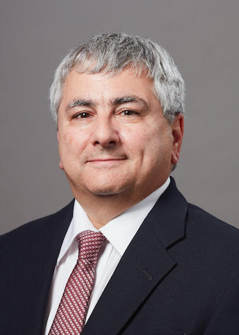
Q: Patient-reported outcomes (PROs) are health care outcomes, such as symptoms or quality of life, reported directly by a patient. In recent years, PROs have emerged as a potentially powerful new way to understand cancer outcomes. Could PROs lead to the next breakthrough in our understanding of cancer?
A: When biochemist and Nobel Prize winner Kary Mullis spoke to my undergraduate class some 20 years ago about his invention of the PCR method for genetic amplification, he put it in historical context by mentioning that every major clinical advance has been preceded by a breakthrough in scientific investigative methods that allowed us to “see” in new ways. Dutch scientist Antonie Van Leeuwenhoek’s microscope allowed scientists to see cells for the first time, advancing us beyond the notion of cancer being caused by an abundance of black bile. The PCR method allowed us to see and manipulate cancer at the genetic level, leading us down the road to targeted therapies aimed at specific genetic mutations. Advances in computer hardware and modeling techniques have allowed us to map the genomes of cancers, moving us beyond a simplistic organ-based model of disease and setting up the possibility of new drug discoveries in silico. Observing and understanding how cancer cells interact with circulatory and immune systems led to VEGF inhibitors, PD-1/PD-L1 inhibitors, and the list goes on…
As our understanding of cancer grows to include larger and more complex systems, we should start to pay serious attention to how cancer interacts with systems outside the human body; its ecology, so to speak. In the last decade cancer researchers have drawn inspiration from studying how invasive species, such as the zebra mussel, take root and overwhelm the ecology of native systems. The word “ecology” comes from the Greek oikos, meaning “home” or “place to live,” and relates cancer to its organic and inorganic environment; not just within the host, but the environment of the host itself. We’ve known for some time how specific exposures to pathogens and substances can influence the rates of specific cancers and how diet, activity, and social network can in some cases influence cancer rates and outcomes.
However, through smartphones, wearables, and integrated sensors, tools to capture PROs are becoming more mainstream and sophisticated in their ability to record granular details about a person’s environment, routine, exposure, and more. Such advancements may be opening up a new era where these details become as important to prognosis and treatment decision making as clinical metrics like cancer stage and genetic profile. Just as we are entering a new era of cancer disease classification based not on primary location but on genetic profile and targeted therapy response, the next sea change may be classifying cancers by their relation and responsiveness to environmental variables and self-care behaviors. Human behavior is complex, but if privacy and regulatory considerations can be worked out and financial incentives aligned, the technology and tools exist to understand it and cancer in a way that’s only been hinted at in epidemiology and health services research up to now.
Copyright: This is an open-access article distributed under the terms of the Creative Commons Attribution License, which permits unrestricted use, distribution, and reproduction in any medium, provided the original author and source are credited.

https://lundberginstitute.org/wp-content/uploads/2018/01/cdg-speech-bubble-trimmed-300-1.png 300 300 melindaroberts https://lundberginstitute.org/wp-content/uploads/2020/08/Artboard-1.png melindaroberts2018-09-04 17:07:242018-09-04 17:07:24Patient-Reported Outcomes Could Transform Cancer Care
Curious Dr. George | Plumbing the Core and Nibbling at the Margins of Cancer

Cancer Pain and the Opioid Epidemic

July 23, 2018/in CDG at TLI


A Q&A with Kevin Sevarino, MD, PhD, President-elect of the American Academy of Addiction Psychiatry and Consulting Psychiatrist at Gaylord Hospital in Wallingford, CT
Q: Opioid abuse, addiction, and overdose are huge American problems right now. Many cancer patients experience chronic pain. What is the best way to use opioids to manage chronic pain?
[Note: The views expressed below represent the opinion of the author, and do not necessarily reflect the views of the American Academy of Addiction Psychiatry nor those of Gaylord Hospital.]
A. We live in amazing times. Targeted immunotherapies, stem cell transplants of transfected cells, identification of unique molecular targets in cancer cells through differential gene expression profiling—all promise to expand survival rates (or cures!) with diminished adverse effects compared to the “blunt hammer” approach of chemotherapy, radiation treatments, and more.

Kevin Sevarino, MD, PhD


But for cancer survivors today, of which it is estimated there are over 14 million in the United States, some 40% live with chronic pain. The pain may be the result of residual effects of the cancer or its treatment, recurrence of the cancer, or development of a new cancer, as well as the many other causes of chronic pain that also afflict those without cancer, such as degenerative joint disease. This pain often goes untreated, since reporting the pain may represent cancer recurrence to the patient, and providers may take a nihilistic view of the pain. Some 25% of patients dying of cancer are in pain.
The opioid epidemic that exploded on the American consciousness has brought to light the role inappropriate opioid prescribing played in triggering the crisis. To reverse harm by the medical profession, organizations like the American Pain Society and the American Academy of Pain Medicine have released guidelines since the late 2000s that emphasize four key points for use of opioids in the treatment of chronic pain in “non-cancer” patients: 1) aim for both adequate pain control and functional improvement; 2) preferentially use non-pharmacologic treatments via multidisciplinary teams and non-opioid pharmacological management, and then if needed, the judicious use of opioids when alternative therapies prove inadequate; 3) maintain high vigilance for the development of opioid use disorders, including use of “universal precautions”; and 4) discontinue opioids in the face of severe adverse reactions. It was not until these were represented as Centers for Disease Control and Prevention (CDC) guidelines in 2016 that widespread attention was paid, as before then the guidelines had little effect on opioid prescribing practices.
So what happens to the cancer patient or survivor who has chronic pain—do the guidelines not apply? Among the few guidelines addressing chronic pain management in cancer patients and survivors, those released by the American Society of Clinical Oncology in 2016 reaffirmed most of the points raised for “non-cancer” chronic pain. Cancer patients face unique pain conditions, including bony metastases, post-chemotherapy pain syndromes, functional loss, and severe psychosocial stresses that have led medical professionals to feel opioids may be better indicated for them than others. But can we say these differ significantly from the stressors faced by those with severe chronic obstructive pulmonary disease, debilitating cardiovascular disease, post-stroke para- or quadraplegia and neuropathy, and more? Yet, the medical field tends not to treat them the same.
I believe there are unique aspects of chronic pain in cancer survivors, but our ability to extend life in the face of terminal illness makes such distinctions increasingly artificial. Our goal should be to extend quality life for as long as possible, and that includes maximizing function, in all people. If one applies the first principal of aiming for adequate pain control balanced with improved function, we should be able to apply the CDC guidelines to cancer patients and survivors not in palliative care or in active cancer treatment.
I am struck by leading cancer treatment center guidances and literature reviews published as recently as 2017 that continue to say opioid use in the treatment of cancer pain rarely results in addiction. This is frighteningly close to what was asserted for opioid use in the treatment of chronic non-cancer pain only 15 years earlier, a bombshell misperception whose splash partially caused the opioid tsunami.
Is the risk of an opioid use disorder lower in the cancer patient with chronic pain, or do we choose to not recognize it or just accept it in the face of a diagnosis so loaded with emotional reaction? Working in a hospital specializing in the care of those with traumatic brain injury and spinal cord injury, I know of the devastating effects lowered expectations can have on treatment outcomes and conversely, the power of positive thinking. If opioids are provided with minimal restriction, and assessment of adverse consequences is not performed, how would a developing addiction be recognized?
If one’s days are numbered by a terminal illness such as cancer we should aim to make those years as meaningful as possible, and the human condition demands those years have meaning; without functioning, without being needed, almost no one feels fulfilled. So, personally, I throw out the contention that the cancer survivor has a lower threshold than those without cancer for the use of opioids when they cannot be shown to be effective for pain control or that they do not cause harm, such as a reduced level of function (interaction with family, continuing to work, etc.). Harm does include ignoring or undertreating pain, but that does not equate to turning to opioids for pain management when so many other modalities may be of benefit. Harm in this population includes the development of an opioid use disorder as well as the indirect provision of opioids to others, either through patient agreement or theft.
Pain management is complicated, it is multimodal, it is time consuming, it is emotionally draining at times, and it is not done well today. What has been learned in oncology, addiction psychiatry and medicine, and other specialties can inform as well as be informed by what we have learned in the rest of medicine.
Copyright: This is an open-access article distributed under the terms of the Creative Commons Attribution License, which permits unrestricted use, distribution, and reproduction in any medium, provided the original author and source are credited.

https://lundberginstitute.org/wp-content/uploads/2018/01/cdg-speech-bubble-trimmed-300-1.png 300 300 melindaroberts https://lundberginstitute.org/wp-content/uploads/2020/08/Artboard-1.png melindaroberts2018-07-23 09:56:342018-07-23 09:56:34Cancer Pain and the Opioid Epidemic
Curious Dr. George | Plumbing the Core and Nibbling at the Margins of Cancer

How to Tell a Patient Their Cancer Has Spread

June 25, 2018/in CDG at TLI

A Q&A with crisis communication expert Lisa Dinhofer, MA, CT

Q. As a counselor and communicator, you are expert and experienced in managing serious situational difficulties up to and including coping with sudden unexpected death. How would you think it best to approach a person with cancer who is being told, “your cancer has spread”?
A: I’ll answer this question by posing another—how did you discuss the diagnosis initially? Did you jointly establish expectations for addressing this illness going forward?
How a diagnosis is delivered plays a critical role in future conversations around how the illness is responding—or not—to treatment. This initial conversation is the foundation for many more that could go in various directions dependent on disease progression, regression, and patient tolerance.
It’s about process and setting the expectation that you are partnering with the patient in their care, which will include honest and compassionate discussion about options as they become available or diminish. How individuals view a diagnosis changes over time. What can’t be imagined initially may become preferred eventually. Leave room for the unknown.
Initial communication principles that include, “As we address this illness, as we see how your illness is responding, we can continue to make decisions based on what we’re seeing,” set a stage for gentle openers and segues if the need to relay unwanted news becomes necessary. Referencing the illness’s response versus the patient’s, “failure” to respond to treatment rests on the disease not the person.
Strive for balance between optimism, hope, and acknowledgement of the situation’s seriousness. Hope and honesty are not binary. Neither are pragmatism and sensitivity. When allowed, hope’s definition can change in meaning resonant with fluid situations.
A talented artist friend battling lung cancer that had spread to her brain remarked that “hope had become a leash” used by family to drag her from coping and conversing honestly in a way she so desperately needed and wanted in her remaining time. She became more prolific as her illness progressed, enough for a successful gallery show, and used her work to “break through” to her family. Her hope transformed from being cured to preparing her young daughter and husband for what lay ahead. We met in pottery class where she made the urn for her cremains.
The following phrasing suggestions incorporate points above with basics for giving bad news:

  1. “(Patient’s name), we need to discuss your latest test results. Honestly, they are disappointing.” (Pause). This is a “warning shot,” giving the patient an opportunity to psychologically “suit up.”
  2. “The tests reveal the illness has spread to ________. (Pause for a few beats to sink in. Rushing on increases the likelihood they won’t hear anything else.) I’m so sorry, (name.)” (This is an apology for their circumstances, not your failure).
  3. “What this means is _______________.”
  4. “Here are options for us to consider_____________.”

If a terminal condition, that does not mean there are no options; it means there are different options than before. The goals of your care might change from treatment to palliative, dependent on a patient’s perspective.
The most important principles for delivering difficult news are preparation, controlling beforehand any personal discomfort so as to completely focus on them rather than rushing to end the conversation, telling what you know when it is known to be true, and remembering that this is about them, not you.
Copyright: This is an open-access article distributed under the terms of the Creative Commons Attribution License, which permits unrestricted use, distribution, and reproduction in any medium, provided the original author and source are credited.
 

https://lundberginstitute.org/wp-content/uploads/2018/01/cdg-speech-bubble-trimmed-300-1.png 300 300 melindaroberts https://lundberginstitute.org/wp-content/uploads/2020/08/Artboard-1.png melindaroberts2018-06-25 09:51:442018-06-25 09:51:44How to Tell a Patient Their Cancer Has Spread
Curious Dr. George | Plumbing the Core and Nibbling at the Margins of Cancer

Cancer Staging: Important Work of the AJCC

January 10, 2018/in CDG at TLI

Jeffrey E. Gershenwald, MD, FACS, Dr. John M. Skibbter Professor, Dept. of Surgical Oncology; Professor, Department of Cancer Biology; Medical Director, Melanoma and Skin Center, The University of Texas MD Anderson Cancer Center, Houston, TX; Member, American Joint Committee on Cancer (AJCC) Executive Committee; Member, AJCC Eighth Edition Editorial Board (Melanoma Expert Panel & Content Harmonization Core); Email: jgershen@mdanderson.org

Q: Staging of cancer is traditionally important. What is the AJCC, who funds it, what does it do? How essential is it for the proper care of cancer patients?

A: The American Joint Committee on Cancer (AJCC) is a multidisciplinary organization with roots dating back to 1959 with the goal of developing and promoting national standards for cancer staging in the U.S. Currently, 22 member organizations spanning multiple disciplines – cancer care, registry, cancer control, pathology, epidemiology, education, outreach and advocacy – support its mission. The AJCC provides worldwide leadership in the development, promotion and maintenance of evidence-based systems for the classification and management of cancer in collaboration with multidisciplinary organizations dedicated to cancer surveillance and to improving care. Administrated by the American College of Surgeons (ACoS) with governance overseen by representatives from founding and other sponsoring organizations, financial support is also provided by several of its member organizations. Notably, other than its core administrative team, it is a nearly completely volunteer-led organization that receives evidence-based input from hundreds of expert volunteers.
Cancer staging serves as the principal communication tool between physicians and their patients and among physicians for clinical decision making and prognostic assessment. Cancer staging is also used for clinical trial design, eligibility, stratification, and analysis. It serves a critical role as the foundation for reporting on an institutional, regional, state, national, and international registry level to facilitate understanding of the broader cancer landscape. With all of this, cancer staging is a key pillar in translational research.
The AJCC published the First Edition AJCC Cancer Staging Manual in 1977. To maintain relevance, AJCC cancer staging systems have been revised and expanded every 5 to 7 years across myriad disease sites included in the manual. The most recent version – the Eighth Edition AJCC Cancer Staging Manual – was published in late 2016 and implemented nationwide in the U.S. on January 1, 2018. It is likely that other countries will also adopt these new staging systems.
The foundational elements of AJCC staging are disease-site specific and anatomic-based (TNM system). Components of the TNM staging system include T (primary tumor), N (regional node and non-nodal regional disease), and M (distant metastasis). The TNM-based system has been employed globally, and continues to benefit from a 30-year partnership with the Union for International Cancer Control (UICC). Cancer staging is performed for a patient at presentation as well as at varying times during their cancer continuum. Such staging classifications include: clinical and pathological (the two most commonly used), posttherapy/post-neoadjuvant therapy (for patients who receive “upfront” systemic and/or radiation treatment as an initial component of their care), recurrence/retreatment (for formal cancer restaging), and autopsy (aTNM). Patients are grouped into cohorts according to risk into various prognostic stage groups.
Despite its success over the past several decades, TNM/anatomic-based staging systems have been de facto constrained in their ability to accommodate improved understanding of cancer biology. In order to be useful, however, they need to be clinically relevant, reflect contemporary practice, and be optimally refined by iterations as our understanding of a given cancer matures.
In an effort to retain clinical relevance, the AJCC has expanded its principles of cancer staging to include non-anatomic based factors (e.g., Gleason score, PSA, mitotic rate) beginning with the 6th Edition (2002), and in the 8th Edition has integrated molecular signatures into some staging chapters (e.g., breast). Formal AJCC acceptance criteria have also been recently developed to serve as a framework for inclusion of contemporary risk models. These changes are overall reflective of a strategic evolution from population-based staging to a more personalized approach.
Given the rapid advances in our understanding of the clinical, molecular, and immunological underpinnings of cancer across multiple cancer types, it is likely that a less “staccato” approach to cancer staging will be devised and implemented by the AJCC; strategically configured and coordinated iterative or “rolling” updates can more efficiently exploit integration of clinically relevant advances into the cancer staging arena. Failure to maintain relevance in this exciting and unprecedented era of cancer discovery and care will eventually render any staging system obsolete.
Jeffrey Gershenwald’s contact info is included in the author affiliations at the top of this page.
Copyright: This is an open-access article distributed under the terms of the Creative Commons Attribution License, which permits unrestricted use, distribution, and reproduction in any medium, provided the original author and source are credited.
https://lundberginstitute.org/wp-content/uploads/2018/01/cdg-speech-bubble-trimmed-300-1.png 300 300 melindaroberts https://lundberginstitute.org/wp-content/uploads/2020/08/Artboard-1.png melindaroberts2018-01-10 15:51:082018-01-10 15:51:08Cancer Staging: Important Work of the AJCC
Page 12 of 22«‹1011121314›»

We Value Your Privacy


Main Menu

  • Our Mission
  • Dedication
  • Our Platform
  • Who We Are
  • Contact
  • Resources
  • Press
  • Curious Dr. George Blog

Recent Press & Resources

  • What Diversity, Equity, and Inclusion Mean for the Health Sciences, and Why Big Data Needs Them All
  • Using Real World Outcomes from “People Like Me” to Guide Cancer Treatment
  • The Strong Helping Hand of Cancer Commons
  • Seeking Patients with Advanced Cancer for a New Clinical Study
  • Morning Administration of Cancer Immunotherapy May Help Patients Live Twice as Long

This Site is Secure


Scroll to top