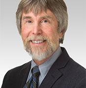
Curious Dr. George | Plumbing the Core and Nibbling at the Margins of Cancer

How Would a Harvard Oncologist Manage His Own Metastatic Kidney Cancer?

Curious Dr. George
Cancer Commons Editor in Chief George Lundberg, MD, is the face and curator of this invitation-only column
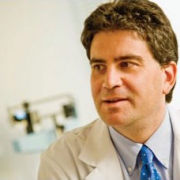
David J. Einstein, MD
Assistant Professor of Medicine, Harvard Medical School & Genitourinary Oncologist, Beth Israel Deaconess Medical Center

When facing a frightening new cancer diagnosis, some people ask their doctors, “What would you do if you were me?” Here, our Curious Dr. George asks kidney cancer expert David J. Einstein, MD, how he would handle his own hypothetical case of kidney cancer. Dr. Einstein is Assistant Professor of Medicine in the Genitourinary Oncology Program at Harvard Medical School’s Beth Israel Deaconess Medical Center.

Curious Dr. George: Please consider this hypothetical scenario—you are an active clinical oncologist in general good health. But lately you have been feeling a little more tired than usual and have lost a bit of weight without dieting, so you decide to visit your primary care physician for a checkup. On physical examination, you are within normal limits, but a routine urinalysis demonstrates microscopic hematuria. A CT scan finds a rounded, 7-cm mass in the upper pole of your left kidney. On chest x-ray, one 2-cm rounded mass is found in the lower lobe of your right lung, and another similar mass is seen in the upper lobe of your left lung. How would you proceed?

David J. Einstein, MD: Before I dive into any of the oncology specifics, I would first acknowledge that this is a very difficult experience and that the most anxiety-provoking time of all is between hearing the diagnosis and learning of a treatment plan. I would definitely want to make sure that I have adequate social supports in place, understand my disability insurance and leave policies, and have access to a dedicated palliative care team to support me physically and emotionally as I embark on cancer-directed treatment.

The scenario outlined here is one of presumed metastatic disease, albeit without widespread metastases. We would first confirm that the pulmonary nodules are in fact metastatic kidney cancer with a biopsy, if feasible. We would then have a multidisciplinary evaluation. Assuming the pulmonary nodules are involved, I would think of this as “oligometastatic,” meaning a gray zone in between widely metastatic and localized.

I would start by assessing symptoms. In this case, it sounds like I have been feeling somewhat unwell, so I do need to start treatment. There are some situations in which we encounter de novo or recurrent metastatic kidney cancers that are asymptomatic and low-volume, and can sometimes be safely watched with close surveillance. We also do a clinical risk stratification. This is mostly for prognostic purposes, although there are some subtle distinctions regarding which systemic therapies are approved for which risk groups.

Then, I would be faced with the decision between systemic therapy first versus some combination of local and systemic therapy. In a situation of more widely metastatic disease, I would certainly opt for systemic therapy first, with some question of coming back to surgery later on, depending on my response and how much disease was within the kidney versus elsewhere.

In this situation, however, it may be reasonable to be aggressive with local therapies, assuming I am in good health. I would consider nephrectomy—removal of the kidney entirely—and some form of local therapy to the lung nodules, either surgery or radiation. By using all of these local therapies, we would be trying to render me disease-free, at least macroscopically. However, I would be concerned that microscopically, there could be some leftover cancer cells that could cause later recurrences. Therefore, I would consider receiving “adjuvant” immunotherapy with the goal of decreasing my risk of recurrence.

We have been using immunotherapies, especially immune checkpoint inhibitors, in metastatic kidney cancer for years. More recently, a study showed benefit for using one year of an immune checkpoint inhibitor, pembrolizumab, after surgery for patients with high-risk localized or oligometastatic kidney cancer, resulting in FDA approval. I would be receiving infusions of this every 3-6 weeks, depending on the dose, and monitoring closely for signs of excess immune activation against my own body, creating essentially an autoimmune condition. The fundamental goal here would be long-term remission, trying to be free of both cancer and treatment, for as long as possible.

Dr. Einstein can be reached at deinstei@bidmc.harvard.edu.


***

Copyright: This is an open-access article distributed under the terms of the Creative Commons Attribution License, which permits unrestricted use, distribution, and reproduction in any medium, provided the original author and source are credited.

Curious Dr. George | Plumbing the Core and Nibbling at the Margins of Cancer

How an Expert Would Manage His Own Stage 4 Pancreatic Cancer: An Update

Curious Dr. George
Cancer Commons Editor in Chief George Lundberg, MD, is the face and curator of this invitation-only column

John H. Strickler, MD
Associate Professor of Medicine, Division of Medical Oncology, Duke University and Co-Leader of Duke Molecular Tumor Board

When confronted with a new cancer diagnosis, some people ask their doctors, “What would you do if you were me?” Previously, our Curious Dr. George asked John H. Strickler, MD, how he would handle his own hypothetical diagnosis of metastatic pancreatic cancer. Now, Dr. Strickler provides an updated answer, outlining new options for the nearly 95% of pancreatic cancer patients with KRAS mutations. Dr. Strickler is Associate Professor of Medicine in the Division of Medical Oncology at Duke University and Co-Leader of the Duke Molecular Tumor Board.

Curious Dr. George: Cancer Commons provides information about options to patients with advanced cancer, usually beyond standard of curative care. As an experienced academic and practicing clinical oncologist at Duke University, you have particular interest, training, and experience in gastrointestinal cancer.  What would you do if you personally were discovered to have an asymptomatic, unsuspected, ductal adenocarcinoma of the tail of the pancreas that had already metastasized to your liver?

John H. Strickler, MD: Pancreatic cancer remains one of the most lethal malignancies. Advancements in chemotherapy, immunotherapy, and targeted therapies have helped countless people with advanced solid tumors, but for pancreatic cancer most of these advancements have not made a meaningful impact. However, despite the grim survival statistics and poor prognosis associated with this disease, there is reason to have hope. With improvements in supportive care, chemotherapy, and molecular diagnostics, some patients are living longer and living better. While we have a long way to go, finally progress is being made.

If I were diagnosed with metastatic (stage 4) pancreatic adenocarcinoma, the first thing I would do is find an experienced multi-disciplinary team. This team would give me the best outcomes possible. Members of this team would include experts on the following fields:

Medical Oncology: Although the primary function of the medical oncologist is to provide chemotherapy, typically he or she formulates the treatment plan and coordinates care. In my hypothetical case, the medical oncologist would recommend either gemcitabine alone, gemcitabine with nab-paclitaxel, or FOLFIRINOX. All of these treatments would be reasonable, but combination chemotherapy (gemcitabine + nab-paclitaxel or FOLFIRINOX) would offer the greatest disease control and longest survival.

Additionally, the medical oncologist would be responsible for ordering next-generation sequencing on my tumor tissue to determine if my tumor harbors an “actionable” genetic alteration. Although these actionable genetic alterations are rare, they may make me eligible for immunotherapy or other targeted therapies. Nearly 90% of patients with pancreatic cancer have a KRAS mutation, and advancements in drug chemistry are finally making KRAS “druggable.” KRASG12C mutations are now considered treatable with highly selective KRAS inhibitors, such as sotorasib or adagrasib. Inhibitors of other more common KRAS mutations are now entering the clinic. We are all hoping to see more breakthroughs.

Genetic counseling: Approximately 5% of all patients with pancreatic cancer have a germline (hereditary) mutation in BRCA1/2 or PALB2, and these hereditary mutations predict benefit from platinum-based chemotherapy and PARP inhibitors. Other rare germline mutations can also predispose a patient to pancreatic cancer. Current national guidelines advise germline testing in all patients diagnosed with pancreatic cancer, regardless of family history. Genetic counseling is advised for any patient who tests positive for a pathogenic germline mutation or has a strong family history.

Palliative care: Many patients are hesitant to consider palliative care. There is a misconception that palliative care represents end-of-life care. I hope that we can change this misconception. Pancreatic cancer often presents with complicated symptoms that are difficult to manage. Even if I present completely pain free, symptoms from pancreatic cancer can change rapidly. Given the incurable nature of this disease, it is helpful to have a team of doctors who can help me and my family prepare for the future. I view palliative care as a critical “extra layer of support” to fight a very difficult illness.

Other important members of the team: As a medical oncologist, I have learned that I am only as good as the people around me. I cannot overstate the importance of having experienced and dedicated nurses, advanced practice providers (NPs and PAs), and clinic staff to provide extra support. Additionally, by finding a skilled multi-disciplinary team for my hypothetical diagnosis, I would have access to other experts, including radiologists, pathologists, surgeons, radiation oncologists, and gastroenterologists. All of these physicians would be key to my health and symptom management.

Final thoughts:

As a gastrointestinal medical oncologist, I have seen how difficult pancreatic cancer can be for patients and their loved ones. This remains a disease with high symptom burden and poor outcomes. If I were facing this disease myself, I would recognize that it takes a community of dedicated clinicians to keep me living longer and living better. As a patient, I would make myself available to clinical trials and other research. It is through these research efforts that we will change the course of this terrible disease and improve outcomes.

More details about ways to support pancreatic cancer research and resources for patients and families fighting this disease can be found at the Pancreatic Cancer Action Network.

Dr. Strickler can be reached at john.strickler@duke.edu.

Curious Dr. George | Plumbing the Core and Nibbling at the Margins of Cancer

How Would a Northwestern Professor Manage His Own Liver Cancer?

Curious Dr. George
Cancer Commons Editor in Chief George Lundberg, MD, is the face and curator of this invitation-only column

Robert E. Matthews
President and CEO, MediSync (Cincinnati, Ohio)

When facing a frightening new cancer diagnosis, some people ask their doctors, “What would you do if you were me?” Here, our Curious Dr. George asks liver cancer expert Al B. Benson III, MD, FACP, FACCC, FASCO, how he would handle his own hypothetical case of liver cancer. Dr. Benson is Professor of Medicine and Associate Director for Cooperative Groups at the Robert H. Lurie Comprehensive Cancer Center of Northwestern University.

Curious Dr. George: Please consider this hypothetical scenario: As a busy academic oncology practitioner, you know that you contracted hepatitis C many years ago and developed chronic active hepatitis leading to fibrosis before you received sufficient Solvadi to eradicate the virus. The fibrosis led to early cirrhosis, which is now being mo

nitored by your physician. You have experienced recent weight loss and some right upper abdominal pain. Abdominal ultrasound has shown many liver nodules thought to be reactive proliferation. But your most recent serum alpha fetoprotein (AFP) has elevated to 375 ng/ml, raising the suspicion that you may now have hepatocellular carcinoma (HCC), perhaps multifocal. How would you proceed?

Al B. Benson, MD, FACP, FACCC, FASCO: I would certainly be concerned about now having a diagnosis of HCC with the known risk of having had hepatitis C, evidence of cirrhosis, and development of some symptoms. Having said that, I would recognize that the current AFP result is a bit controversial since acute inflammation and liver cell regeneration can cause an increase in AFP. It would appear that the abdominal ultrasound suggests reactive proliferation without definite evidence of HCC. Since I would be worried that the standard screening with AFP and ultrasound in my case may not have definitely ruled out HCC, I would favor proceeding with additional imaging, such as multiphasic CT scans. Once I had a contrast-enhanced abdominal CT scan I would then request review by a multidisciplinary team. If the team—particularly from the viewpoint of radiology, interventional radiology, and hepatology—agree that the lesions do not fit the criteria for HCC and are confident a biopsy would not be indicated, I would continue monitoring with very close follow-up. It would be important to make sure my liver function tests (LFTs) were not changing in terms of my cirrhosis, and there would need to be discussion as to the etiology of my symptoms.

Let us assume, however, that my CT scan fits imaging criteria for HCC. Of note. if imaging criteria are met, a biopsy would not be necessary for diagnosis, although I might still consider a biopsy—particularly for analysis by next-generation sequencing (NGS)—for the chance that there might be a molecular profile which could inform a therapeutic choice or participation in a clinical trial. If imaging was inconclusive, I would favor a biopsy to confirm the diagnosis.

Prior to a biopsy, I would want review by a transplant team as part of a multidisciplinary discussion to determine if I was a potential resection or transplant candidate, which for HCC would be my preferred treatment choice if I met the criteria for either resection or transplantation. If it was clear that resection or transplantation was not feasible, and there was no evidence of metastatic disease on CT imaging—including chest, abdomen and pelvis—then I would review with interventional radiology about the possibility of liver-directed therapy, such as Y90 radioembolization, including the possibility of participating in an available liver-directed therapy clinical trial.

Systemic therapy would be the choice if liver-directed therapy was not an option. A number of biologically driven therapies have been developed, including immunotherapy, expanding choices for individuals, with additional regimens in development in clinical trials. In my case, I would undergo an esophagogastroduodenoscopy (EGD) to evaluate presence/stability of varices, and if there is no contraindication, consider therapy with atezolizumab plus bevacizumab, with tremelimumab plus durvalumab as an alternative choice. If symptom control became an increasing issue, I would seek advice from the palliative care team.

To help friends and family better understand the management of hepatocellular cancer, I would refer them to the National Comprehensive Cancer Network (NCCN) guidelines for patients.

Dr. Benson can be reached at albenson@nm.org.

Curious Dr. George | Plumbing the Core and Nibbling at the Margins of Cancer

Successful Control of High Blood Pressure, a Major Cancer Comorbidity

Curious Dr. George
Cancer Commons Editor in Chief George Lundberg, MD, is the face and curator of this invitation-only column

Robert E. Matthews
President and CEO, MediSync (Cincinnati, Ohio)

People with cancer often face additional health conditions, or “comorbidities,” that complicate treatment. High blood pressure, or hypertension, is a major comorbidity that affects many cancer patients. Our Curious Dr. George asks Robert E. Matthews, President and CEO of MediSync in Cincinnati, Ohio, about how his company’s tools are helping physicians better manage their patients’ blood pressure.

Curious Dr. George: All physicians and medical organizations know that hypertension is a major attributable cause of many serious and fatal illnesses, such as coronary artery heart disease, heart failure, and stroke. As many as 48% (119 million) of American adults have hypertension. The American Heart Association, the American Medical Association, the American College of Cardiology and hundreds of other organizations have set a new target of 130/80 (revised from 140/90) for blood pressure control and have launched a major initiative to achieve such. Yet, the Centers for Disease Control and Prevention (CDC) reports that fewer than 48% of American patients with hypertension meet even the less stringent historical 140/90 goal.

Your group practice, PriMED Physicians in Ohio, using the MedsEngine AI tool from MediSync and the NICaS (non-invasive cardiac system with impedance cardiography) test coupled with solid clinical leadership reports that its cooperating institutions have consistently exceeded 90 or even 95% blood pressure control for more than 10 years. Exemplary.

Your approach to matching the correct drug or drugs and dosages with each patient’s unique blood pressure pathophysiology would seem to be the key. Why does your organization succeed, and how might others follow your lead to success?

Robert E. Matthews: Helping patients with hypertensive diseases (HTN) reduce blood pressure (BP) to 140/90 or lower has been and remains a struggle for physicians. Since 1999, CDC reports have shown that far more than half of all diagnosed patients fail to achieve this level of BP control. Recent studies strongly suggest an even more stringent BP goal of lower than 130/80 would benefit most patients.

PriMED Physicians in Dayton, Ohio, has a consistent record of achieving >90% BP control across all HTN patients with very slight month-to-month variations. PriMED has a database tracking key quality metrics, including BP for every patient at every visit for over 10 years.

In October 2023, 94% of all of PriMED’s HTN patients; 95% of all the patients with both HTN and diabetes and, remarkably, 96% of all African American patients from socio-economically challenged zip codes had a BP of less than 140/90. These outcomes reflect a different way of providing chronic disease care. You yourself recently encouraged that, now that we know these results are possible, more groups should be using PrIMED’s methods, which are briefly described here:

  • PriMED formally established a corporate goal of achieving greater than 90% “control” (control is defined as meeting the evidence-based standard definition of control) for several major chronic diseases in 2002 via a groupwide vote of all physicians. Hypertension was the first disease tackled.
  • After surveying non-healthcare companies noted for their consistent quality and discovering that all of them adopted a quality science approach, PriMED and its management partner, MediSync, underwent almost a year of extensive training in the Six Sigma quality science, tools, and statistics.
  • Six Sigma instructs that great care be taken to enumerate the challenges to achieving target goals and, further, to discover the root causes of those obstacles. PriMED and MediSync inquired extensively into the challenges of getting 90% of all HTN patients to the target BP. PriMED’s “root cause analysis” of the problems that result in sub-optimal outcomes is enumerated in this talk co-sponsored by The Lundberg Institute and the Commonwealth Club of California.
  • Most medical groups conclude that “the patient is the problem” when treating hypertension. PriMED and MediSync recognized that the patient is the smallest part of the challenge. The actions of physicians and other providers are key to success.
  • Simplistically described, elevated blood pressure can be the result of several hemodynamic factors, including (1) vasoconstriction, (2) elevated heart rate, (3) elevated stroke index, reflecting a more violent contraction, (4) excessive fluid status, and (5) some combination of 1 through 4. When there are multiple causes, calculating the portion of each cause is very helpful.
  • PriMED’s physicians determined that using a simple, Food and Drug Administration-approved, in-office test called Impedance Cardiography (ICG) provides sufficiently accurate analysis of the hemodynamics behind elevated blood pressure. Statistical analysis demonstrated that, when PriMED’s providers used ICG, their outcomes were significantly improved. Without ICG outcomes were worse.
  • Over time, PriMED recognized 28 additional demographic and comorbid conditions that can influence provider choices for optimal pharmacotherapy by patient. Given the variables described above, PriMED’s calculations include 50 key variables which, in turn, generate over 240 million permutations.
  • All quality sciences prove that doing complex work successfully requires a carefully constructed process. A process is a series of steps designed to achieve the target outcome. If different actors do their work in an individualized manner, quality will be significantly lower.
  • PriMED and MediSync first created a “paper and pencil” process to guide providers to select the optimal drugs from the 13 different classes of anti-hypertensive drugs for each patient.
  • In 2017, MediSync made available and PriMED adopted an IT application called the MedsEngine that pulls vast amounts of data from any patient’s electronic health record and identifies and calculates the variables to make individualized drug recommendations for the patient. Provider use of this application is highly correlated to outcome success.
  • The MedsEngine permitted PriMED to automate more disease calculations for cholesterol, diabetes, and heart failure—diseases for which the group also has excellent outcomes especially by national standards.

Medicine in general and primary care in particular have yet to broadly adopt or embrace the quality improvement techniques and methods that are ubiquitous in virtually all other economic sectors. It is clearly possible and urgently important that primary care does a far better job getting chronic diseases controlled. Once a few medical groups accomplish remarkable goals, there is no further debate about whether it is possible. Hundreds of millions of patients can live longer, healthier lives on average if we meet the challenge of better outcomes.

Mr. Matthews can be reached at bob.matthews@medisync.com.

***

Copyright: This is an open-access article distributed under the terms of the Creative Commons Attribution License, which permits unrestricted use, distribution, and reproduction in any medium, provided the original author and source are credited.

Curious Dr. George | Plumbing the Core and Nibbling at the Margins of Cancer

High-Energy Blue Light Powers a Promising New Treatment to Destroy Cancer Cells

Curious Dr. George
Cancer Commons Editor in Chief George Lundberg, MD, is the face and curator of this invitation-only column

Stuart L. Marcus, MD, PhD
Founder & Chief Science Officer, SonALAsense

A perennial challenge in cancer treatment is figuring out how to kill tumor cells while leaving healthy cells unharmed. Our Curious Dr. George asks Stuart L. Marcus, MD, PhD, founder and Chief Science Officer at SonALAsense, how his company’s light-activated drug treatment addresses this challenge, holding potential promise for people with brain, bladder, and possibly other cancers.

Curious Dr. George: Your flagship treatment employs sonodynamic therapy (SDT), in which focused ultrasound is used to generate a light that, enabled by a metabolite of aminolevulinic acid (ALA) concentrated selectively in cancer cells, activates the oxidative destruction of those cancer cells but not other cells. How does that work? And what can you tell us about how effective it is for cancer patients?

Stuart L. Marcus, MD, PhD: To answer how SDT works, we have to first understand that SDT is a new, noninvasive form of a light-activated drug therapy called ALA photodynamic therapy (PDT). ALA PDT targets the heme pathway selectively in cancers. When provided with excess ALA, the fluorescent photosensitizer protoporphyrin IX (PpIX) accumulates primarily in cancer tissue to such a specific extent that the U.S. Food and Drug Administration (FDA) has approved ALA-induced fluorescence in a number of different cancer settings. For instance, urologists use it to help identify bladder cancers through blue light cystoscopy. Neurosurgeons also use it every day as a visual aid to resect high-grade brain cancers, such as glioblastoma multiforme (GBM), employing blue light resection and fluorescence-detecting surgical microscopes.

When PpIX is exposed to the high energies of blue light, energy is transferred from PpIX to molecular oxygen (the photodynamic process), making it highly reactive. In 1999, the FDA approved a topical ALA PDT blue light system, which I and my team at DUSA Pharmaceuticals created for treatment of the precancerous skin condition actinic keratosis. That system, now marketed by Sun Pharmaceutical Industries, has treated over 4 million people to date. ALA PDT has also been successfully used in investigational clinical studies to treat recurrent GBM (rGBM) using the ingested dye Gleolan as the source of ALA and by inserting fiber optics within the tumor which deliver laser light. Although I considered further developing PDT for rGBM, this system is so invasive and complex that, in my opinion, commercialization is moot as the treatment is very difficult to standardize.

Instead, with ALA SDT, we use MR-guided focused ultrasound to safely create light in tissue through a process called sonoluminescence. In preclinical studies, we and others have shown that this approach can effectively and selectively kill gliomas in animal models through the PDT process and boost the animals’ survival. In a first-in-man energy dose-ranging study at the Ivy Brain Tumor Center, each of 10 rGBM patients received our intravenous form of ALA, which is called SONALA-001, and we applied SDT to one-half their tumor. The patients’ tumors were surgically removed in 4 days, and oxidative changes (lipoperoxide liberation) and apoptosis were compared in the treated and untreated halves of each tumor. We found that ALA SDT selectively caused PDT oxidative damage to the treated tumor half.

We are now carrying out a therapeutic phase 2 dose-ranging clinical trial in rGBM patients. We are also treating children with the deadly cancer diffuse intrinsic pontine glioma (DIPG), in which we treat the entire brain stem. We have seen no dose-limiting toxicities as yet in either trial, attesting to the tumor-specificity of ALA. The heme pathway is a universal pathway, and the tumor-specific PpIX accumulation response to being provided with excess ALA gives us hope that SDT will prove therapeutic for many types of tumors.

Dr. Marcus can be reached at smarcus@sonalasense.com.

***

Copyright: This is an open-access article distributed under the terms of the Creative Commons Attribution License, which permits unrestricted use, distribution, and reproduction in any medium, provided the original author and source are credited.

Curious Dr. George | Plumbing the Core and Nibbling at the Margins of Cancer

Integrating AI-Generated Learning Materials into Health Professional Education

Curious Dr. George
Cancer Commons Editor in Chief George Lundberg, MD, is the face and curator of this invitation-only column

Miriam Chickering, RN, BSN, NE-BC
CEO of NextGenU.org

Artificial intelligence (A.I.) is a powerful tool that has made its way into diverse fields—and medical education is no exception. Our Curious Dr. George asks Miriam Chickering, RN, BSN, NE-BC, CEO of NextGenU.org, how the 22-year-old, U.S.-based organization she leads uses A.I. to help create free educational materials for healthcare professionals and students.

Curious Dr. George: NextGenU.org provides teaching materials and curricula in a wide range of medical, public health, and humanitarian fields for more than 2,000 institutions and countless students in all countries, free of charge. With such a vast mission, NextGenU.org would seem an ideal organization to utilize large language model-based generative A.I. in creating original course materials. Products such as ChatGPT have taken the information world by storm in recent months. Yet many users have experienced serious problems of facts and misrepresentation in A.I.-generated materials.

What is the scope of use of such products as ChatGPT by NextGenU.org in creating course materials, and what quality control measures do you use to minimize errors?

Miriam Chickering, RN, BSN, NE-BC: Integrating A.I. into health professional education (HPE) holds undeniable promise—a future where boundless, personalized educational resources and experiences redefine how students prepare for the complexities of healthcare. However, this bright future is not without its challenges. The landscape is mired with concerns over the quality of A.I. outputs, which, if not addressed, could compromise the integrity of HPE and the safety of healthcare delivery.

Incorporating A.I. into HPE requires a thoughtful process. NextGenU.org’s current and evolving process is as follows:

  1. Educate the Team on effective interaction with A.I. tools and evaluation of A.I.-generated content.
  2. Evaluate each A.I. Tool beginning with a thorough assessment of the A.I.’s capacity, dataset, and the expertise and philosophy of its developers.
  3. Identify Integration Points to determine areas for A.I. enhancement within each workflow process.
  4. Develop and Test A.I. Prompts to generate specific outputs, and conduct comprehensive testing to optimize the prompt for accuracy.
  5. Curate A.I. Inputs with vetted, high-quality resources to ensure relevant and accurate output generation.
  6. Create Content such as multiple-choice questions and interactive activities while ensuring alignment with educational objectives.
  7. Ensure Quality Control with subject matter experts validating the accuracy and pedagogical soundness of outputs. Experts ensure outputs align with the approved inputs or align the outputs with reliable and relevant resources.
  8. Integrate Content into the curriculum, and review and act upon student feedback while monitoring student outcomes.
  9. Expand Learning Opportunities through the use of A.I., offering infinite, safe chances to practice.
  10. Optimize Resource Allocation by shifting resources saved through A.I. efficiency towards teaching soft skills and nuanced clinical judgment.
  11. Practice Continuous Improvement through monitoring and refining the use of A.I. tools.

Evaluating an A.I. tool is like bringing a new research assistant to a seasoned research team. As with any new addition, we start by scrutinizing the A.I.’s “credentials”—its dataset and developers—much like evaluating a candidate’s CV for their academic lineage and references.

Once we welcome our “digital assistant” to the team, we don’t entrust it with independent research or content creation. Instead, we provide carefully vetted inputs and detailed prompts, akin to giving a new research assistant specific literature to review or data to analyze. These controlled inputs help ensure that the A.I.’s outputs are standardized. The content is then subjected to a rigorous quality control process. Subject matter experts, akin to senior researchers, oversee and verify the A.I.’s work at each point when the A.I. is used.

We use A.I. assistance for a number of tasks, including:

  1. Compare and contrast competency sets and course structures
  2. Develop objectives and outcomes
  3. Draft multiple choice questions and case studies
  4. Identify web-based learning resources
  5. Summarize learning resources

Readers interested in learning more can watch our videos that demo how we use A.I. assistance to create multiple choice questions and activities. I have also coauthored an academic paper on the use of A.I. in health professional education, and I will participate in a live discussion of this topic in an upcoming webinar on Dec 1.

We envision a future where learners benefit from a virtually limitless sandbox to practice, experiment, and learn from an endless variety of situations, ensuring that when they face real-life challenges, they are well-prepared and confident.

Miriam Chickering can be reached at mchickering@nextgenu.org.

***

Copyright: This is an open-access article distributed under the terms of the Creative Commons Attribution License, which permits unrestricted use, distribution, and reproduction in any medium, provided the original author and source are credited.

How Would an Expert Manage His Own Acute Myelogenous Leukemia?

Curious Dr. George
Cancer Commons Editor in Chief George Lundberg, MD, is the face and curator of this invitation-only column

Adam Asch, MD
Professor of Medicine; Nancy Johnson Records Chair of Oncology, University of Oklahoma College of Medicine

When facing a frightening new cancer diagnosis, some people ask their doctors, “What would you do if you were me?” Here, our Curious Dr. George asks leukemia expert Adam Asch, MD, how he would handle his own case of acute myelogenous leukemia. Dr. Asch is Professor of Medicine and Nancy Johnson Records Chair of Oncology at the University of Oklahoma College of Medicine.

Curious Dr. George: Please consider this hypothetical scenario: You are an active clinical oncologist in general good health. But lately you have lacked energy, have been feeling a little more tired than usual, have some dizziness on exertion, have been bruising easily, and have lost a bit of weight without dieting. So, you decide to visit your primary care physician for a checkup. On physical examination, she notes that you are a little pale, finds arm bruises, and feels the tip of your spleen. She orders several lab tests. Your hematocrit is 38, hemoglobin 12, white blood cells (WBCs) are 49,000 per microliter with many blasts, many promyelocytes, myelocytes, eosinophils, and basophils. Platelets are low. A diagnosis of acute myelogenous leukemia (AML) is made. How would you proceed?

Adam Asch, MD: This would be a fascinating case to discuss in the abstract. But in the first person, as you have been exploring in your Curious Dr. George series, diagnosis and therapy take on a special and sometimes personal urgency. For this 70-year-old oncologist, the hypothetical is a timely one and for the field a relevant one, since AML is, in so many ways, a disease of aging.

For most AML patients, I find that the first, most important question to ask is if there is a curative intervention. For some, chemotherapy induction and consolidation may lead to a prolonged remission and perhaps cure. But for some disease categories—notoriously, p53-mutated disease—response to traditional chemo is poor. Second, assuming an initial remission follows an induction regimen, how a remission is consolidated is another critical question. For many people with high-risk AML (complex cytogenetics, FLT3-mutated) as well as for intermediate-risk disease, allogeneic transplant remains the best means of consolidating a remission and offering cure.

To a great extent, treatment for AML is informed by genetic and molecular studies that have become standard in the field. Cytogenetics to assess chromosomal defects, molecular analysis to identify potentially targetable mutations, and multi-channel flow cytometry to identify a blast phenotype that can be followed to assess measurable residual disease post induction and consolidation are all standard. Translocation 15;17 or “acute promyelocytic leukemia” (APL)—once considered to be the worst type of AML prognostically—is now routinely treated with all-trans retinoic acid and arsenic trioxide, with long-term survival now approaching 90%. The clinical presentation in my hypothetical case does not sound like APL, though the bruising raises the question, and the coagulation profile, if indicative of disseminated intravascular coagulation, might make that more of a question. Other good prognostic cytogenetics are translocations of 8;21 and inversion ch16. Each of these has a historical long-term survival of about 60 to 70% with standard induction chemotherapy (I’ll say another word about this in a bit). All other cytogenetic features are classified as intermediate risk or poor risk, with long term survival rates of 30-40% or 10%, respectively. For each of these latter risk groups, transplant remains the only real chance of relapse-free, long-term survival.

Now, before we discuss treatment regimens for AML in general, there are some features of my hypothetical presentation that would lead me to ask for some additional data. The presence of a spleen tip and the presence of early myeloid forms and eosinophils and basophils in addition to blasts raises the possibility that my disease is a Ph+ blast crisis rather than de novo AML. If so, treatment with a kinase inhibitor directed at the BCR-ABL kinase—after initial cytoreduction of the elevated blast count—would be a reasonable initial approach to getting control of the disease. But without allogeneic transplant as consolidation, this would not offer me long-term control of the disease. So, more on transplant in a bit.

Standard induction for “fit” patients with AML remained largely unchanged for several decades—until recently. The chemo combination of 7+3 (7days of cytosine arabinoside and 3 days of an anthracycline) has been the backbone in the U.S. of induction. Consolidation chemotherapy with 3 to 4 subsequent cycles of high-dose cytosine arabinoside has been common practice. The development of a liposomal preparation containing a defined ratio of cytosine arabinoside and daunorubicin has shown increased efficacy in AML that arises from a pre-existing condition known as myelodysplastic syndrome (MDS). Also, drugs targeting specific mutations in genes such as FLT3, IDH1, or IDH2 are additional options that can now be used in combination with standard approaches—or alone in some instances. But perhaps most impressive has been the incorporation of the BCL2 inhibitor venetoclax in combination with azacytidine as a low-intensity regimen now approved by the U.S. Food and Drug Administration (FDA) for elderly and unfit patients with MDS. Response rates appear essentially equal to those of the standard high-intensity induction regimens. Importantly, this regimen generally has lower toxicity and is most often administered outpatient. Recent data shows that patients achieving a complete response with this regimen do as well with allogeneic transplant as folks who received more intensive induction. And for younger, fit patients, venetoclax added to more intensive regimens is producing results that are likely significant improvements over the old 7+3.

So, getting back to the first question I posed, am a looking at the possibility of a cure? Possibly. It is not likely, at my age, that cytogenetics would be favorable. Complex cytogenetics and AML arising from MDS are much more prevalent. Even if this were to be a Ph+ AML blast crisis, my long-term survival would depend on allogeneic transplant as consolidation post remission.

Am I a candidate at 70 for an allogeneic transplant? Possibly. Recent studies show that arbitrary age limits on allotransplantation have virtually disappeared across transplant centers. Less ablative regimens and improved supportive care around infection control and prevention of graft-versus-host disease have led to a situation where transplantation for older patients is a viable option with reasonable outcomes. Unfortunately, it is one that is currently afforded to only about 5% of older AML patients.

So, what would I want in this situation? For my intermediate or poor-risk AML, I would likely opt for azacytidine and venetoclax as an induction/consolidation regimen, which would give me a better chance of escaping potentially debilitating complications of induction and consolidation that might put a definitive transplant out of reach. And, I would consider transplant as consolidation if in complete remission and still in good shape.

Some reality here: if these approaches didn’t get me to a complete remission, my discussions would center on defining the best way to preserve quality of life with family and friends during my remaining time.

Dr. Asch can be reached at adam-asch@ouhsc.edu.

***

Copyright: This is an open-access article distributed under the terms of the Creative Commons Attribution License, which permits unrestricted use, distribution, and reproduction in any medium, provided the original author and source are credited.
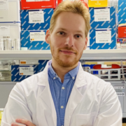
How Would an Expert Manage His Own Advanced Esophageal Cancer?

Curious Dr. George
Cancer Commons Editor in Chief George Lundberg, MD, is the face and curator of this invitation-only column

Sarbajit Mukherjee, MD, MS
Asst. Prof. of Oncology and Co-Leader, GI Translational Research Group—Roswell Park Comprehensive Cancer Center (Buffalo, NY)

When facing a frightening new cancer diagnosis, some people ask their doctors, “What would you do if you were me?” Here, our Curious Dr. George asks gastrointestinal cancer expert Sarbajit Mukherjee, MD, MS, how he would handle his own case of advanced esophageal cancer. Dr. Mukherjee is Assistant Professor in the Department of Medicine—GI Medical Oncology and Co-Leader of the GI Translational Research Group at Roswell Park Comprehensive Cancer Center in Buffalo, NY.

Curious Dr. George: Imagine a hypothetical scenario in which, as a relatively young clinical oncologist, a lifelong non-user of tobacco, and not an alcohol abuser, you did not imagine that you might be afflicted by a gastrointestinal malignancy. You have experienced some epigastric discomfort and have taken antacids from time to time. But recently, sometimes your solid food does not go down as easily as usual, and you have lost five pounds. Your primary care physician suggests esophageal endoscopy, which finds an obstructing distal esophageal mass. Biopsy reveals a poorly differentiated adenocarcinoma, and chest CT shows a widened distal esophagus, three suspicious mediastinal lymph nodes, and a single 2 cm pulmonary mass. When you learn of this unfortunate situation, how do you proceed?

Sarbajit Mukherjee, MD, MS: Esophageal cancer is the eighth most common cancer globally. As of 2022, the lifetime risk of developing esophageal cancer was 1 in 125 men and 1 in 417 women for the U.S. population. Recent studies have shown that young-onset esophageal adenocarcinoma (age at diagnosis under 50 years) is rising. Unfortunately, young-onset cases often present at an advanced stage, resulting in poor survival.

Over the last few years, we have had several new approvals in esophageal cancer, but the prognosis for metastatic cancer remains poor. Each patient’s treatment is individualized, and specific information about the tumor’s molecular characteristics helps guide a treatment decision.

Initially, I would complete the staging work-up with a CT scan of the abdomen and pelvis with contrast. I would check for biomarkers, particularly HER2, PD-L1 CPS score, and MSI-H status. I would also consider doing next-generation sequencing testing of the tumor tissue. To confirm the metastatic disease, I recommend a CT-guided biopsy of the pulmonary nodule.

I would ensure that an expert medical oncologist sees me at a tertiary care center. I would also ask my oncologist if any clinical trials are available for me in an upfront setting. If I were not enrolled in a clinical trial, I would receive a 5-fluorouracil-based chemotherapy with or without trastuzumab and an immune-checkpoint inhibitor (pembrolizumab or nivolumab), depending on the results of the biomarker status. Before starting any treatment, I would want to ensure my nutritional status is adequate and see a nutritionist.

There are several other factors to consider while caring for a young-onset esophageal cancer patient. Firstly, symptom management during cancer therapy is extremely important. Therefore, I would seek a referral to palliative care. I would also ensure a living will or an advanced directive is in place. Research has shown that early interdisciplinary supportive care improves survival in metastatic esophageal cancer. A new cancer diagnosis could be devastating for any family, and I would seek a psychology referral to help my family cope with the diagnosis.

Secondly, fertility preservation should be discussed with every cancer patient in the reproductive age group. If the patient wishes to, they should be referred to an oncofertility specialist to discuss some of the established means of fertility preservation before starting systemic treatment.

Finally, I would also seek a genetic counseling referral to determine if my cancer is potentially inheritable so my children can be screened accordingly.

In summary, young-onset esophageal cancer is on the rise and often presents at an advanced stage. Biomarker testing is important for making a personalized treatment plan. Patients should be offered participation in clinical trials whenever possible. Early supportive care, genetic counseling, and fertility preservation should be considered for this unique population.

Dr. Mukherjee can be reached at sarbajit.mukherjee@roswellpark.org.

***

Copyright: This is an open-access article distributed under the terms of the Creative Commons Attribution License, which permits unrestricted use, distribution, and reproduction in any medium, provided the original author and source are credited.

Alibrex: A New Blood Test to Reveal Whether a Cancer Treatment is Working

Curious Dr. George
Cancer Commons Editor in Chief George Lundberg, MD, is the face and curator of this invitation-only column

Kevin Knopf, MD
Chief Medical Officer, Cadex Genomics; CMO and Division Chief of Hematology/Oncology, Highland Hospital (Oakland)

In cancer treatment, time is of the essence. Our Curious Dr. George asks Kevin B. Knopf, MD, MPH, Chief Medical Officer at Cadex Genomics, how his company’s new test, Alibrex, could rapidly reveal how well a given treatment is working for a given patient.

Curious Dr. George: The U.S. Food and Drug Administration has approved about 270 anti-cancer drugs. The National Comprehensive Cancer Network publishes guidelines for treatment of about 61 major cancer types. Although there is consensus on first- and second-line therapy, there may be a substantial delay before a patient’s oncologist can determine whether the therapy chosen is working for them. Your company Cadex Genomics has a new test to evaluate whether an agent is active against a tumor in a given patient. You claim that your test, Alibrex, is agnostic to tumor type, tissue of origin, and the name of the drug and can give an answer quickly. Can you describe the test, how it works, and how it is performing at this early time?

Kevin B. Knopf, MD, MPH: Alibrex is being developed to identify stage IV cancer patients who are not responding to treatment, allowing for an early switch to more effective therapy. Specifically, the test identifies patients whose tumor is rapidly growing during treatment. Alibrex requires a blood draw prior to therapy being administered, and another prior to the next cycle of treatment. Using quantitative polymerase chain reaction qPCR technology, cell-free DNA (cfDNA) concentration levels are measured in each blood draw to determine if the tumor is growing.

The concept is straightforward: as tumors grow, the amount of cfDNA the tumor sheds into the blood increases. Alibrex takes advantage of this by picking up the increase in cfDNA and determining whether the patient has disease progression. The advantage of evaluating cfDNA versus circulating tumor DNA (ctDNA) is that all tumors shed cfDNA with increasing amounts as the tumor grows. Compared to cfDNA, ctDNA is a far more complex approach in that different tumors shed different types of ctDNA.

A concern with this approach is that increases in cfDNA may be seen from other non-cancer-related sources, such as infections, white blood cell lysis, injury, or a number of other events unrelated to tumor growth. It is possible for a patient with such an event, prior to a blood draw, to have a result that is a false positive or negative due to the impact of changing levels of non-cancer cfDNA.

To overcome this problem, Alibrex assesses short cfDNA fragment lengths; it has been published and recognized that short cfDNA fragments are specific to cancer. By focusing on short cfDNA fragments, Cadex Genomics has developed a tumor-agnostic biomarker that is uniquely fit for purpose as a therapy monitoring tool.

To be able to detect small changes in cfDNA, Alibrex must be highly reproducible and have very low limits of detection to meet the performance necessary for clinical utility. We achieve this by targeting a non-coding region of DNA that has 1,800 copies in every cell and is located across all 23 chromosomes. Alibrex can detect differences in cfDNA levels in less than what is in a single cell.

Alibrex has been analyzed in two patient cohorts. The first, a study of 32 stage IV colorectal cancer patients at MD Anderson Cancer Center, demonstrated a sensitivity of 63% and specificity of 100%. A later multi-center study (74 lung, breast, and colorectal cancer patients) on an optimized version of the assay, demonstrated that Alibrex has a sensitivity of 88% and a specificity of 100%. By the end of August of 2023, we will have expanded this 74-patient analysis to 150 patients. Prior to making the test available to patients in the clinical setting, we will validate the assay in a 100-patient lung cancer study.

In clinical practice, we anticipate Alibrex being used monthly to determine if a patient is developing progressive disease and not responding to therapy. We do not anticipate Alibrex to be an imaging replacement; rather, we see the potential of imaging being used in significantly longer intervals.

In the future, with additional studies, Alibrex may potentially be used to monitor therapy in the neoadjuvant setting and to monitor prostate cancer patients who are in active surveillance.

Dr. Knopf is also Chief Medical Officer and Division Chief of Hematology and Oncology at Highland Hospital in Oakland, CA. He can be reached at kevinbknopf@gmail.com.


***

Copyright: This is an open-access article distributed under the terms of the Creative Commons Attribution License, which permits unrestricted use, distribution, and reproduction in any medium, provided the original author and source are credited.


Common Sense Oncology: Moving Back Towards Outcomes that Matter

Curious Dr. George
Cancer Commons Editor in Chief George Lundberg, MD, is the face and curator of this invitation-only column

Christopher M. Booth, MD, FRCPC
Medical Oncologist, Queen’s University, Kingston, Canada; Co-Founder, Common Sense Oncology

There are many ways to measure how well a particular cancer treatment works. But how much do these outcomes reflect a patient’s actual wellbeing? Our Curious Dr. George asks Christopher M. Booth, MD, FRCPC, one of the founding leaders of the new international initiative “Common Sense Oncology” (CSO), how CSO is working to improve patient outcomes.

Curious Dr. George: The practice of clinical oncology is difficult, at best, dealing every day with patients with potentially lethal malignancies. Some segments of oncology seem to have lost their way, veering towards the money-driven medicine that tempts physicians in many countries. How does CSO endeavor to put the patient at the forefront in the drive to maximize survival and enhance quality of life in an equitable manner, whilst giving serious attention to cost?

Christopher M. Booth MD, FRCPC: The art of oncology involves compassionate care, clear communication, and treatment that offers meaningful benefits.

Over the last four decades, transformative advances in surgery, radiotherapy, and systemic therapy have led to improved survival and quality of life for patients. However, in the last decade, a worrisome trend has emerged: most new cancer medicines do not help people live longer or better lives. Rather, the majority of new treatments approved by the U.S. Food and Drug Administration (FDA) are based on delaying tumour growth on a CT scan—usually measured as progression-free survival (PFS). In some circumstances, improvements in PFS are known to translate into improved overall survival (OS); however, these are the exception rather than the rule.

Hence, contemporary oncology is faced with a major challenge: most of our older treatments improve OS and/or quality of life, but most new medicines do not. This worrisome trend in our field is not well appreciated by patients or policymakers because our field has been consumed by hype—even for treatments that only have small benefits. The recent shift in our field to new medicines with marginal benefits is important because these treatments come with substantial costs: clinical toxicity for patients, time toxicity for patients and families, and financial toxicity to patients, families, and the health system.

These problems have developed due to a complex convergence of hope and money. Physicians and patients inherently look for treatment to provide benefit in what can be an overwhelming emotional journey. In many circumstances, a natural tension arises between hope and reality; this tension is most acute in the context of incurable cancer. In parallel, massive financial forces are involved in modern cancer medicine. These financial forces shape the research agenda and delivery of care. The pharmaceutical industry now funds about 90% of cancer drug trials, and a substantial proportion of industry revenues are directed into mass-marketing campaigns designed to increase use of cancer therapies—even if they are associated with marginal benefits.

In response to these challenges, an international group of oncologists, academics, and patient advocates gathered at Queen’s University in Kingston, Canada, in April 2023 to launch CSO. This grassroots initiative seeks to ensure all patients have access to cancer treatments that improve outcomes that matter, such as OS and quality of life. CSO’s mission, vision, guiding principles and action plan were recently published in the academic journal Lancet Oncology and on the Common Sense Oncology website. CSO is an inclusive movement that welcomes engagement from all stakeholders in the global cancer system. The voices of patients and patient advocates have been a core component of CSO since its inception. CSO promotes informed decision making with patients and seeks to promote health justice through access to high-quality and affordable cancer care.

The vision of CSO is that patients have access to cancer treatments that provide meaningful improvements in outcomes that matter, irrespective of where they live or their health system. To realize this vision, CSO has launched three streams of work related to evidence generation, evidence interpretation, and evidence communication.

Through our evidence generation efforts, CSO will work to ensure that clinical trials measure and report the impact of new therapies on outcomes that matter to patients. Evidence interpretation will involve educational initiatives to increase critical appraisal and critical thinking skills among oncologists. Our evidence communication initiatives will seek to improve understanding of cancer treatment risks and benefits among patients, the public, the media, and policymakers.

One of the primary goals in launching CSO was to provide a forum for the oncology community to have candid conversations about the successes and failures within our field.  CSO will celebrate well-conducted trials and treatments that measurably improve the lives of patients. However, the CSO community will also be willing to call out treatments that may cause more harm than good. Efforts to improve the generation, interpretation, and communication of evidence in our field will help move oncology closer to a reality in which all patients have access to high-quality treatment with meaningful benefits—regardless of where they live.

Dr. Booth is a medical oncologist and health services researcher at Queen’s University in Canada, where he holds the Canada Research Chair in Population Cancer Care. He can be reached at christopher.booth@kingstonhsc.ca.

***

Copyright: This is an open-access article distributed under the terms of the Creative Commons Attribution License, which permits unrestricted use, distribution, and reproduction in any medium, provided the original author and source are credited.